Cavus Foot in Children: When Should We Be Concerned?

Dieser Artikel ist nur auf Englisch verfügbar.
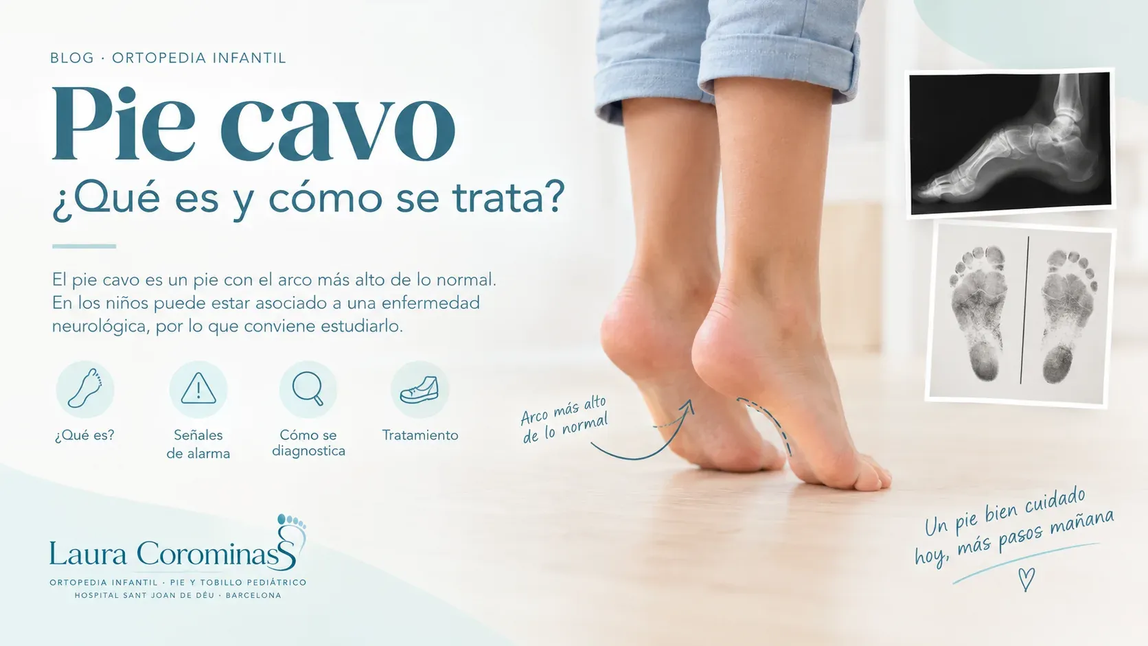
Have you noticed that your child has an unusually high arch, that their shoes wear unevenly, or that they frequently twist their ankle? They may have a cavus foot.
In some children, this foot shape causes no discomfort and requires little more than routine monitoring. However, during childhood, it is important to assess why it develops, particularly if the deformity progresses over time or is associated with pain, falls, or recurrent ankle sprains.
What is a cavus foot?
A cavus foot is a deformity in which the arch of the foot is higher than normal. It is often associated with an inward tilt of the heel, known as hindfoot varus, and clawing of the toes.
This combination can lead to an uneven distribution of body weight, increasing pressure on the heel and the forefoot.
Not all cavus feet are the same. Some are flexible and partially correct when the foot position changes, while others are more rigid and may significantly affect gait.
How can we recognise it?
Here are some signs that parents and caregivers may notice:
-
An unusually high arch, even when the child is standing.
-
Claw toes, particularly when associated with other deformities.
-
Increased weight-bearing through the heel and forefoot, with reduced contact through the midfoot.
-
Calluses or areas of thickened skin at pressure points.
-
Recurrent ankle sprains or a feeling of instability.
-
Pain when walking, running, or playing sports.
-
Fatigue, frequent tripping, or difficulty finding comfortable footwear.
A high arch alone does not necessarily indicate an underlying condition. What matters is assessing the shape of the foot, its flexibility, any associated symptoms, and how it changes as the child grows.
Why does cavus foot develop?
This is one of the most important questions.
In some children, cavus foot has a familial component and remains stable for years. In others, it may be associated with an underlying neurological or neuromuscular disorder that causes an imbalance between the muscles of the foot and ankle.
Possible causes include:
-
Neuromuscular disorders, such as Charcot-Marie-Tooth disease.
-
Neurological or spinal cord abnormalities.
-
Certain genetic disorders or syndromes.
-
Familial cases in which no associated disease is identified.
For this reason, when assessing a child with cavus foot, we should look beyond the shape of the arch. It is also important to examine muscle strength, reflexes, sensation, and gait.
Particular attention should be paid if the cavus deformity develops or worsens over time, affects one foot more than the other, or is associated with muscle weakness, sensory changes, or balance difficulties.
How is cavus foot assessed?
Assessment begins with a comprehensive clinical examination.
During the consultation, we evaluate the shape of the foot and heel, toe position, flexibility of the deformity, ankle range of motion, and the strength of the different muscle groups. We also assess the child’s gait and look for signs of an underlying neurological condition.
Depending on the clinical findings, we may request:
-
Weight-bearing X-rays: to assess bone alignment and the severity of the deformity.
-
Neurological assessment: if findings suggest an underlying neuromuscular disorder.
-
Additional investigations: such as electromyography, genetic testing, or MRI, when indicated.
Not every child needs all these investigations. The diagnostic work-up is tailored to the clinical examination and the progression of each individual case.
How is cavus foot treated?
Treatment depends on three key factors: the underlying cause, the flexibility of the foot, and the symptoms it produces.
1. When the foot is flexible and painless
When the deformity is mild, stable, and does not limit activity, clinical follow-up may be sufficient.
Not every cavus foot requires orthotic insoles or surgery. The aim is to monitor its progression and identify any changes early.
2. When it causes pain or difficulty walking
In symptomatic cases, conservative measures may be considered, including:
-
Comfortable footwear with sufficient room for the toes.
-
Custom orthotic insoles to improve pressure distribution and foot support.
-
Physiotherapy and tailored exercises, according to the child’s needs.
-
Treatment of ankle instability, when present.
Orthotic insoles can help relieve symptoms, but they do not necessarily correct an established structural bone deformity.
3. When the deformity is significant or progressive
When cavus foot is rigid, causes persistent pain, interferes with walking, or leads to recurrent instability, surgical intervention may need to be considered.
Depending on the type of deformity, surgery may involve osteotomies — procedures that correct bone alignment — soft-tissue procedures, or tendon transfers to improve muscle balance.
In certain neuromuscular conditions, addressing the underlying cause and planning long-term follow-up are also essential.
The surgical approach is selected on an individual basis: there is no single procedure suitable for every cavus foot.
Can cavus foot worsen during growth?
Yes, in some cases. This is why follow-up is important, particularly when the deformity is progressive or associated with an underlying neuromuscular disorder.
A stable, flexible, asymptomatic cavus foot is not the same as one in which the arch becomes increasingly elevated, claw toes develop, or pain and recurrent ankle sprains begin to occur.
Identifying these changes allows us to investigate the underlying cause and determine the most appropriate treatment at each stage of growth.
When should you consult a specialist?
I recommend seeking an assessment if you notice that your child:
-
Has a very high arch with the heel tilting inward.
-
Experiences recurrent ankle sprains.
-
Develops pain, calluses, or difficulty participating in sports.
-
Has claw toes or trips frequently.
-
Develops a deformity that progresses over time.
-
Has muscle weakness, sensory changes, or a family history of neuromuscular disease.
If the deformity progresses rapidly or new muscle weakness develops, prompt medical assessment is advisable.
Dieser Artikel dient der allgemeinen Information und ersetzt keine fachärztliche Untersuchung.